History of Anatomical Dissection

Why is dissection important?

Dissection of the human body is the only method of direct observation and measurement of the structures, organs, bones, ligaments and tendons that allow the body to function. Dissection is a fundamental part of the training of physicians and other care providers. Physicians cannot treat disease or trauma without a complete understanding of anatomy.

Timeline

History of Anatomical Dissection

Fifth-Sixth Century BC

Hippocrates and his followers make first detailed records of human dissections. They established a rational scientific, approach to the treatment of disease. Laws against dissection were inscribed on stelae on Hippocrates’ home island of Cos. Plato’s discussion of human anatomy was based solely upon religious belief. 
Third Century BC

Greek physicians Herophilus of Chalcedon and Erasistratus of Chios perform human dissections in Alexandria, Egypt and Antioch.

157 AD

Galen of Pergamum (129-c.216 AD) served as physician to a troop of gladiators maintained by the high priest of Asia; also served as physician to Roman emperor Marcus Aurelius. Roman law prohibited dissection and autopsy of the human body.

162 AD

Galen gave public demonstrations in anatomy; dissected and experimented on African monkeys, sheep, pigs and goats.

Galen identified seven pairs of cranial nerves, described heart valves, and differentiated between veins and arteries; showed that arteries carry blood not air.

Prevailing prohibitions on human dissection forced him to dissect animals and led to errors. Galen’s description of the human uterus was based on that of a dog. 

536

Sergius of Rashayda translates Galen into Syriac.

873

Persian Physician Hunaya Ibn Ishaq translates Galen into Arabic.

1213

Frederik II Emperor of Germany and King of the Two Sicilies insists a human dissection be performed every five years.

1315

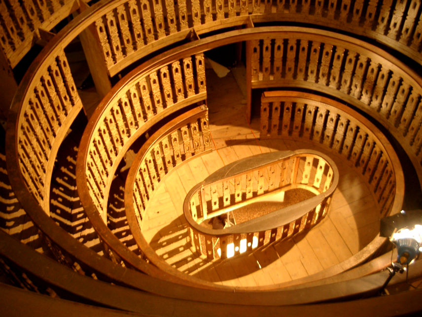
Mondino d’Liuzzi (1270-1326) performs the first officially sanctioned public dissection in Bologna in the presence of medical students and other spectators; responsible for reviving investigation through dissection and inclusion of dissection in the medical curriculum of the University of Bologna.

1316

Mondino wrote his major work, Anathomia corporis humani, considered the first example of a modern dissection manual and the first true anatomical text; in use for next 250 years. 

1490’s

Leonardo Da Vinci did dissections in the hospital of Santa Maria Nuova and broadened his anatomical work into a comprehensive study of the structure and function of the human body; worked on an anatomy text that was never published.

1525

Galen’s works printed in Greek in Europe; Latin translations followed quickly.

1543

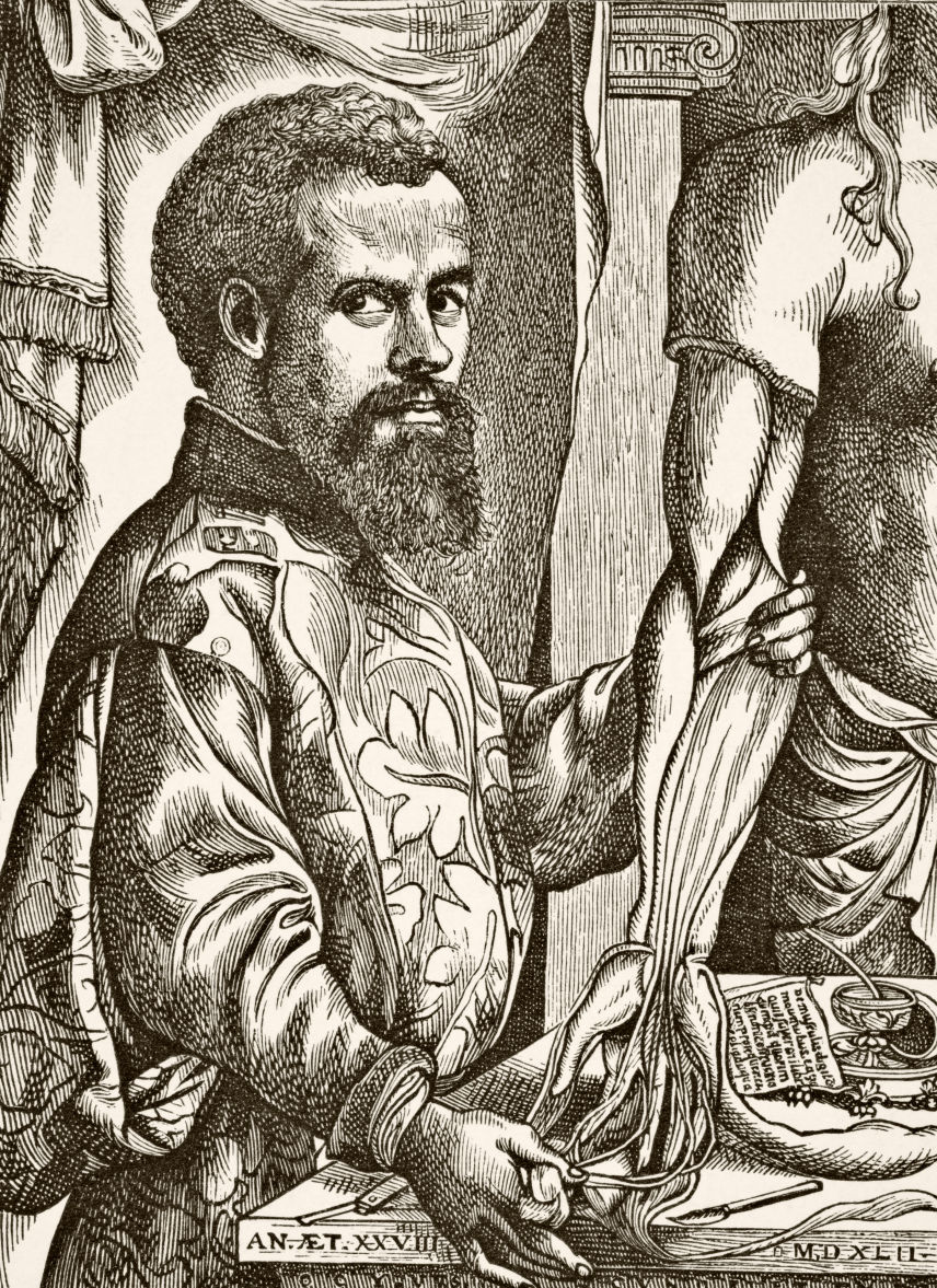
Andreas Vesalius (1512-1564), a Belgian physician, publishes The Seven Books on the Structure of the Human Body (De humani corporis farbrica libri septem).

Based on careful observations from new dissections, this work began to undermine Galen’s authority. Vesalius performed dissections, directed demonstrations, introduced dissection to medical curriculum.

Copernicus published his concept of the heliocentric system.

1628

William Harvey, by observation and reasoning, developed a theory of the circulation of the blood; published his classic work on anatomy and function, Concerning the Motion of the Heart and Blood (Exercitatio Anatomica de Motu Cordis et Sanguinis in Animallibus).

1752

Murder Act in Great Britain allowed the bodies of executed murderers to be dissected for anatomical research and education.

Mid-18th Century

In England, the Royal College of Physicians and the Company of Barber Surgeons were the only organizations permitted to carry out dissections.

1761

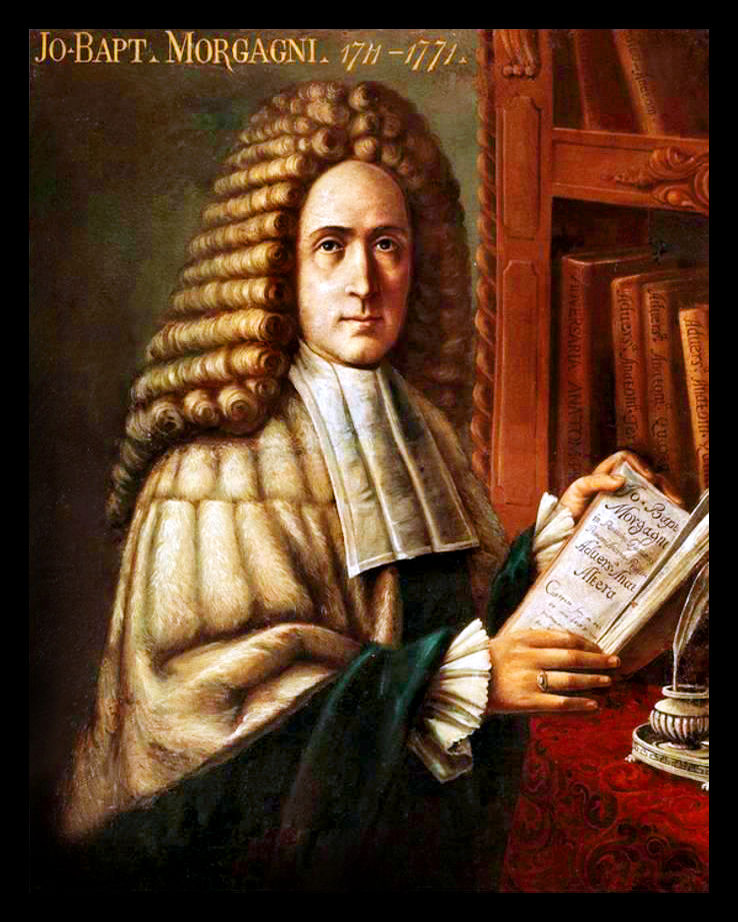
Italian anatomist Giovanni Battisti Morgagni published The Seats and Causes of Diseases Investigated by Anatomy (De Sedibus et Causis Morborum per Anatomen Indagatis). The work contained records of 640 dissections.

A widely respected physician, Morgagni was the first to show the necessity of basing diagnosis and treatment on a comprehensive knowledge of anatomy; considered the father of modern autopsy.

1832

Anatomy Act in the United Kingdom allowed physicians and surgeons to have legal access to corpses that were unclaimed after death, particularly those who died in prison or workhouse. A person could donate a next of kin’s corpse in exchange for burial at the expense of the person accepting the body.

1858

Henry Gray published first edition of his Anatomy: Descriptive and Surgical, later known simply as Gray’s Anatomy. The work covered 750 pages and contained 363 figures. The success of the book was due to the excellence of its illustrations. A second edition was prepared by Gray and published in 1860. The 40th edition was published in 2008.

1885

Illinois Cadaver Act passed. The Act requires that bodies to be buried at public expense be transferred to physicians, schools of medicine or of mortuary science, or other institutes of higher learning for research and educational purposes. It is currently in force.

1910

The Flexner Report, Medical Education in the United States and Canada, dissection becomes an established requirement of the medical school curriculum.

1918

Forerunner of AGA is established in Chicago as the Demonstrators Society. It is intended to accept donations and to provide cadavers to the city’s medical schools and other institutions.

1956

Ultrasound first used for clinical reasons. A Glasgow obstetrician, Ian Donald, and an engineer, Tom Brown, developed the prototype system for clinical use of ultrasound, which was not perfected until the end of the late 1950’s.

1968

National Conference of the Commissioners on Uniform State Laws approved Uniform Anatomical Gift Act. The model law provided that a donor’s intent regarding disposition of his/her body supersedes those of next of kin. It made willed-body programs based on voluntary donations more systematic. By 1972, 48 states had enacted laws similar to/based upon the Uniform Anatomical Gift Act.


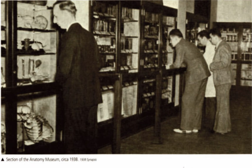
"Image detail of "Section of the Anatomy Museum, circa 1938"
DO from the Philadelphia College of Osteopathy, Class of 1938
Philadelphia College of Osteopathic Medicine Archives, Philadelphia, Pennsylvania"

1972

CT (Computed Axial Tomography) invented in 1972 by British engineer Godfrey Hounsfield and by South African-born physicist Allan Cormack of Tufts University. Anatomy instructor use CT scans to identify and illuminate anatomic structures.

1977

On July 3, 1977, the first magnetic resonance imaging (MRI) exam on a live human patient was performed. Like CT scans, MRIs reveal parts of the body difficult to access.

1990’s

New preservation techniques are developed, such as plastination and latex injection, to preserve anatomical specimens for much longer periods of time.

1992

Thiel method of embalming perfected. This “soft-fix” embalming method preserves the natural look and feel of a cadaver. Limbs, skin and muscles remain flexible. Internal organs are clearly identifiable and respond to the surgeon’s scalpel as if alive.

1994

The National Library of Medicine (NLM) within the National Institutes of Health uses images for clinical medicine and biomedical research. The Visible Human Project was designed to build a digital image library of volumetric data representing complete, normal adult male and female anatomy.

1994-95 The National Library of Medicine releases the Visible man and Visible woman, 3-D computerized cadavers made from compiled MRI, X-ray and photo images of cross sections of a donated body.

2004

The Human Tissue Act adopted in the United Kingdom. It created a priority for control of disposition of cadavers.

2006

Practicing surgical methods on cadavers legalized in the United Kingdom.

2011

CT scans of donor cadavers are used to teach medical students to detect anatomic anomalies before dissection.

2014

Ultrasound study of cadavers used in training of emergency medicine residents to detect the presence of air or gas in the cavity between the lungs and the chest wall, causing collapse of the lung (pneumothorax).

2015

Techniques for augmenting cadaver-based activities, including body painting, use of 3-D prints and progressive dissection, implemented in medical schools.

2020

A commercial scanning laser Doppler vibrometer used on cadavers to enhance the implementation of electronic devices that provide a sense of sound to profoundly deaf or severely hard-of-hearing (cochlear implants).

Address

1540 S. Ashland Ave., Suite 104
Chicago, IL 60608

Contacts

Email: info@agaillinois.org
Office: (312) 733-5283
Fax: (312) 733-5079